Self-pay (non-insured)
Front tooth implant after a root fracture — same-day extraction, bone graft & implant (#11)
Root fracture of a previously root-canal-treated upper central incisor (#11), crowned more than 30 years ago, with resorption of the labial (front) bone wall — confirmed on 3D CT. At the first examination: labial probing depth 7 mm with bleeding on probing, no mobility. Non-restorable; extraction and replacement indicated. The neighbouring upper lateral incisor (#12) had an ill-fitting crown with decay beneath it.


A front tooth crowned more than 30 years ago suddenly felt as if it had shifted while the patient was eating. A 3D CT scan showed that the root beneath the crown had fractured. Below we explain what a root fracture is, why root-canal-treated teeth are more prone to it, and why an implant — placed on the same day as the extraction, with bone grafting and no incision — was chosen to restore the tooth while preserving the gum.
From a shifting front crown to an implant-supported crown — step by step
Treating dentist: Dr. Ayaka Nakamura, D.D.S.Clinical review: Dr. Ryosuke Murai, D.D.S., M.S.D.Published: Last updated:
Why the patient came in: a front crown that suddenly felt as if it had shifted
The patient, a man in his 60s, had an upper front tooth (upper right central incisor, #11) that had been crowned more than 30 years earlier. While eating, the tooth suddenly felt as if it had shifted, and he came to RODIN.
Even when a front tooth looks almost unchanged, a crown that feels loose or moves when you bite can be a sign of a problem in the root underneath it.
At the first examination, the tooth was not loose, but the pocket on the front (labial) side measured 7 mm and bled on probing. A deep pocket confined to one spot on a single tooth, while the surrounding gum is otherwise shallower, can be a sign of a root fracture, as bacteria travel down along the crack.
A 3D CT scan (cone-beam CT) showed that the root of the tooth had fractured, and that the bone on the front (labial) side of the root had resorbed. A root fracture line can be hard to see on a conventional two-dimensional X-ray, so a 3D scan is useful for this diagnosis.
What is a root fracture?
A root fracture is a crack or split in the part of the tooth that sits inside the gum and bone. A fracture can run along the length of the root (a vertical root fracture) or across it (a horizontal root fracture).
Once a crack reaches the root, bacteria can enter along the fracture line and the bone around the root can gradually break down. Symptoms may be mild at first, so bone loss can progress before the patient notices. In this case, the labial bone had already resorbed.
Depending on how and where the root has split, an attempt to keep the tooth (for example by bonding) is sometimes possible. When the crack is deep and the surrounding bone has been lost, extraction is usually the recommended option.
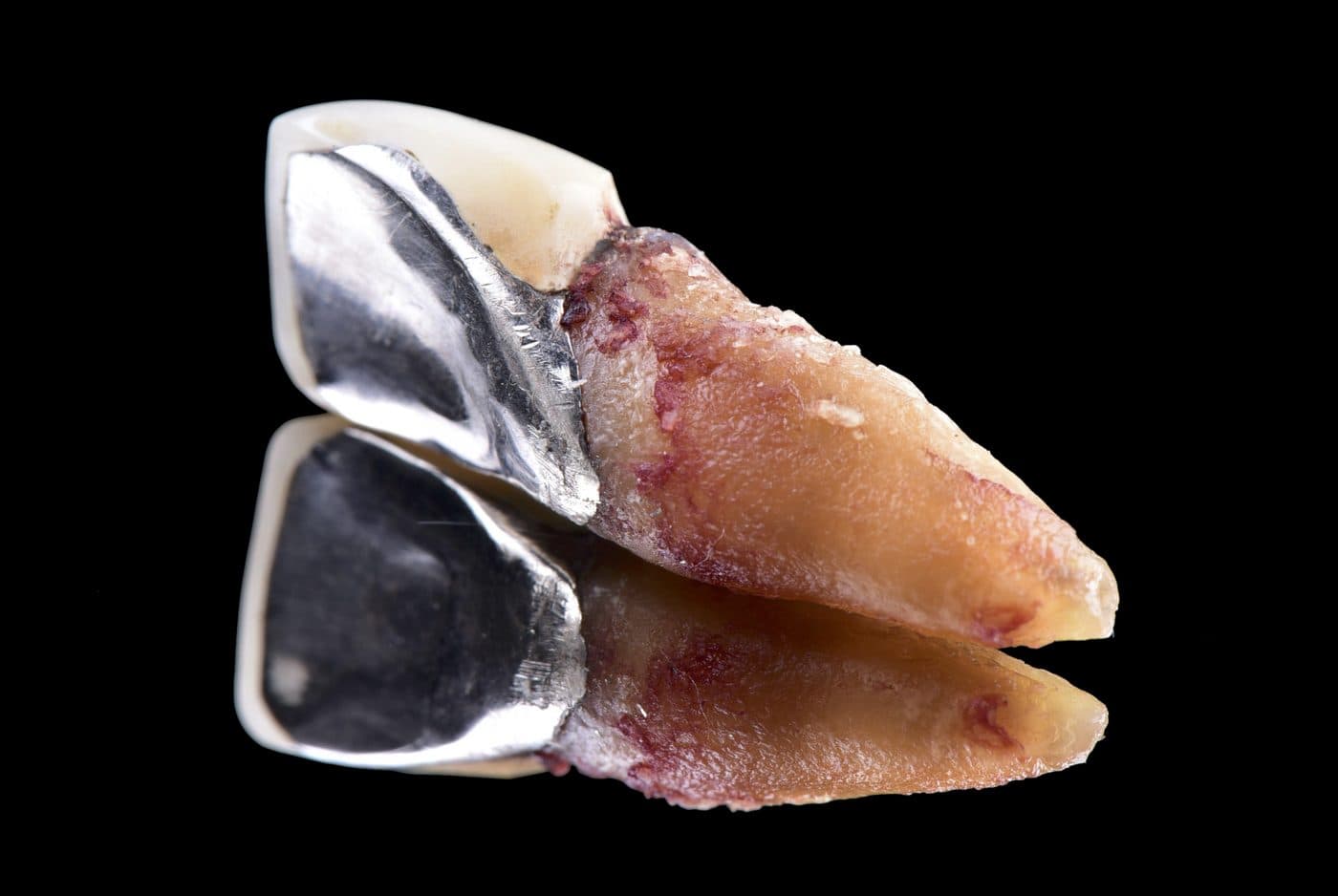
In this case, the extracted tooth showed a crack in the root beneath the crown (photo).
- A crown that feels loose or as if it has shifted
- Pain or discomfort when biting
- Swelling of the gum around the tooth
- A small pimple-like opening on the gum (sinus tract) that releases pus
Why root-canal-treated teeth are more likely to fracture
During root canal treatment, decay and the inside of the tooth are cleaned out, so less tooth structure remains. A post and core are often placed before a crown, and biting forces can concentrate on part of the root. For these reasons, teeth that have had root canal treatment are generally considered to carry a higher risk of root fracture than teeth with a living nerve.
In Japan's nationwide survey of the causes of permanent-tooth extraction (8020 Promotion Foundation, 2nd survey, 2018), fracture accounted for 17.8% of extractions, third after periodontal disease (37.1%) and decay (29.2%). The report states that the proportion of fractures was markedly higher in teeth without a living pulp than in teeth with one.
A crown placed 30 years ago has carried decades of biting force. If a crowned tooth starts to feel different, an early examination is advisable.
Why an implant rather than a bridge
The main options for replacing a single front tooth are an implant, a bridge, or a partial denture. A bridge is attached to the two neighbouring teeth, which are prepared as supports for a joined set of crowns.
In this case, both neighbouring teeth had also had root canal treatment in the past, so they carried the same higher fracture risk. A bridge would have added the biting load of the missing tooth to those teeth, and was considered likely to increase the chance of them fracturing in the same way in future.
The front teeth also have a large impact on appearance. An implant stands on its own without relying on the neighbouring teeth, protecting them while aiming to restore appearance — including the shape of the gum. For these reasons, an implant was chosen.
Extraction, bone grafting and implant placement in a single surgery
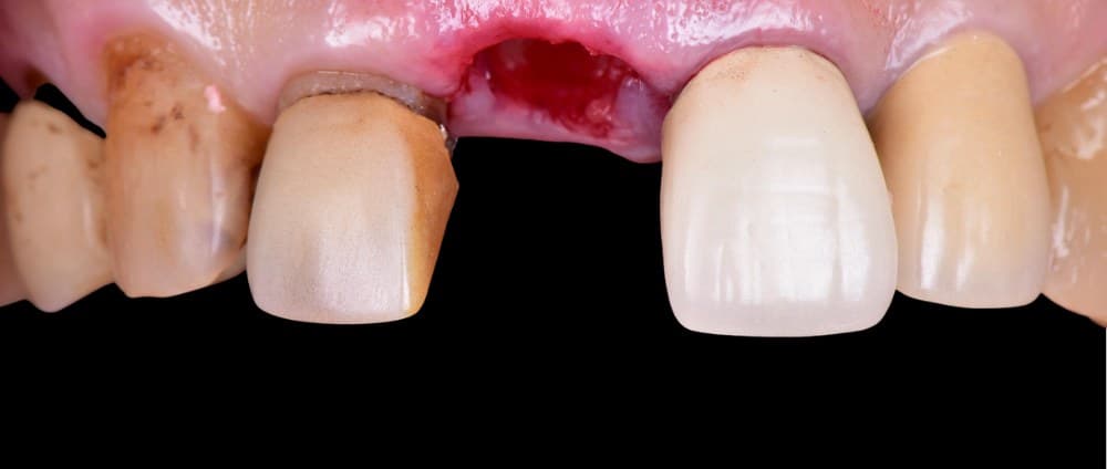
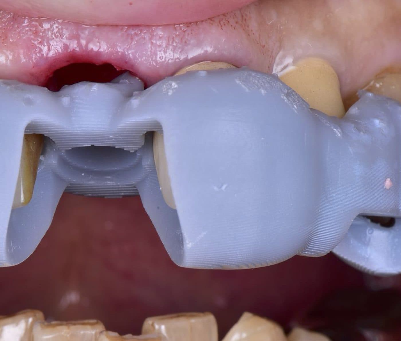
Because the labial bone had resorbed, guided bone regeneration (GBR — rebuilding bone with bone graft material and a membrane) was carried out at the same time as the extraction, and the implant was placed in the same surgery (immediate implant placement).
Bone tends to shrink after a tooth is removed. Combining extraction, grafting and placement keeps it to one surgery. The implant position was planned on the CT scan and transferred to the mouth with a 3D-printed surgical guide. Immediate placement on its own does not prevent this post-extraction bone loss, so bone grafting was combined here to rebuild the labial bone. The ITI notes that a thin facial bone wall resorbs considerably more after immediate placement and that a damaged facial wall is a risk factor for gum recession, so careful assessment on CT — including whether bone grafting is needed — is essential.
Primary stability of 30 Ncm — letting the implant heal without load
How firmly an implant is anchored at the moment of placement (primary stability) is assessed by its insertion torque. With limited bone available, the primary stability in this case was 30 Ncm.
Attaching a temporary crown to an implant and loading it straight away (immediate loading) is considered predictable when conditions are favourable, including sufficient primary stability. Because this case combined bone grafting with modest primary stability, the implant was not loaded, and healing until the implant had integrated with the bone was given priority.
In the meantime, appearance was maintained with a temporary tooth bonded to the neighbouring teeth, so the patient did not have a visible gap at the front during healing — without putting any load on the implant.
No incision and a custom healing abutment to preserve the gum
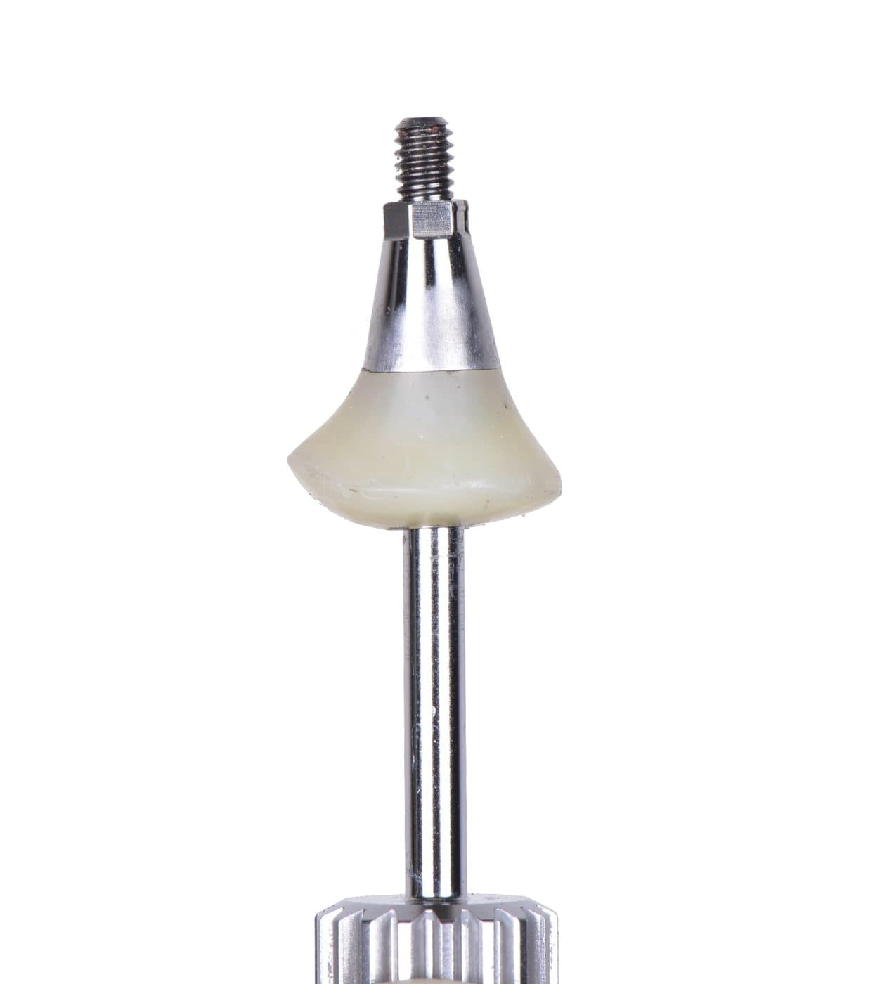
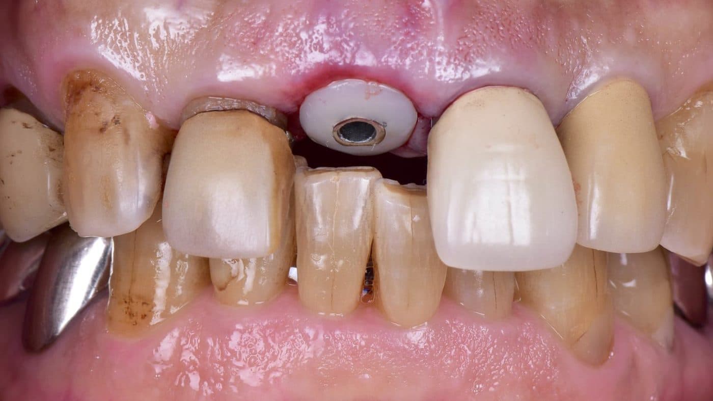
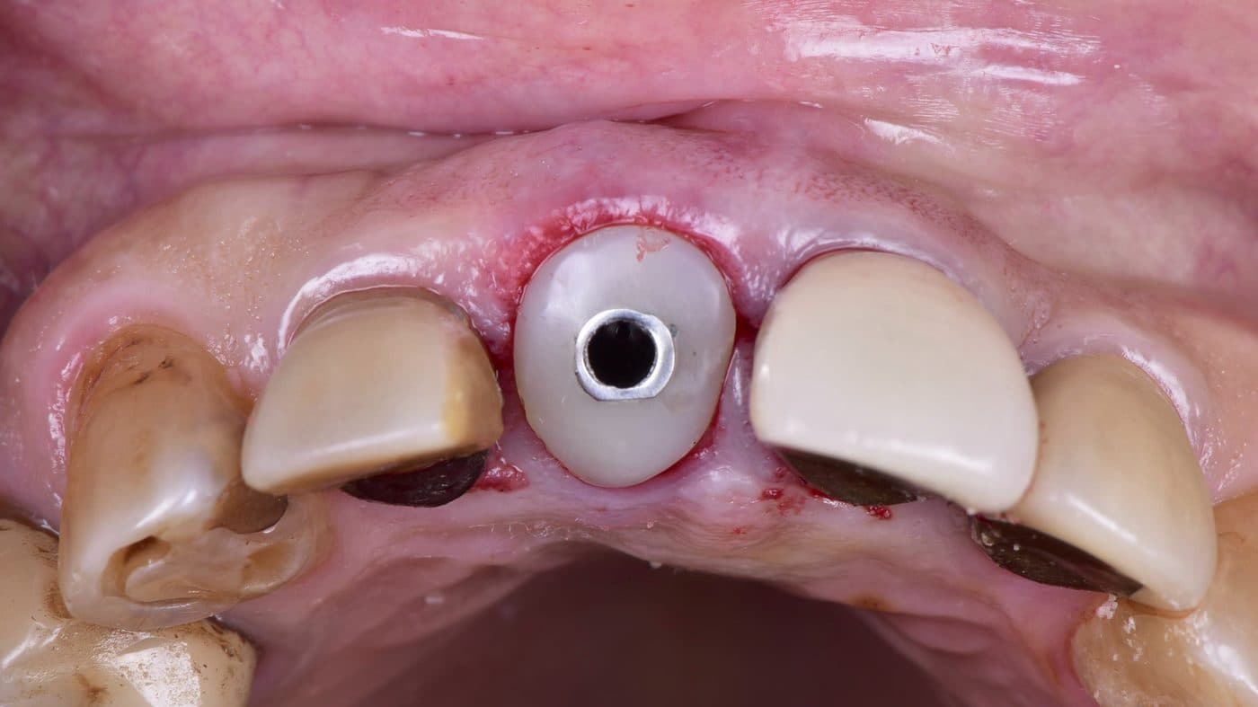
With a front-tooth implant, the shape of the gum matters as much as the bone. To preserve as much gum tissue as possible, no incision was made (flapless surgery), and a custom healing abutment shaped to the contour of the original tooth at the gumline was connected and left in place for about three months.
A custom healing abutment supports the gum from the inside immediately after extraction and guides it towards the emergence shape of the natural tooth. A 2024 systematic review reported that customized healing abutments predictably shape the peri-implant soft tissue, which can improve aesthetic outcomes compared with standard round healing abutments — while noting that the number of studies is small and follow-up periods are short.
In this case, the gum contour was preserved without a gum (soft-tissue) graft (individual results vary).
Checking with a provisional before the final screw-retained zirconia crown
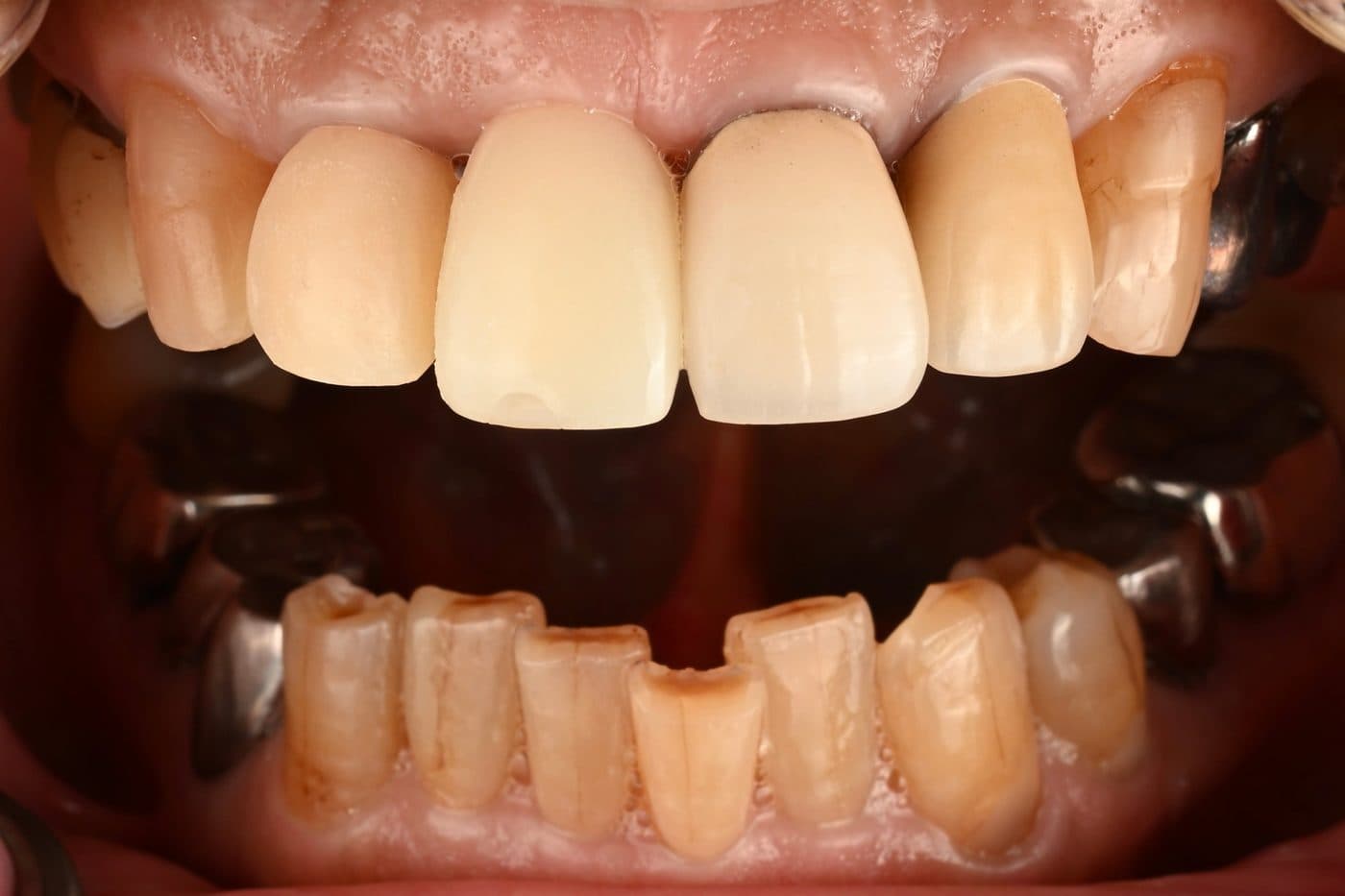
After healing, the bonded temporary was replaced with a provisional restoration connected to the implant. With it, appearance, speech, the bite and the gum contour were checked and confirmed.
The final restoration was a screw-retained zirconia crown. It is fixed with a screw rather than cement, so it can be removed for maintenance if needed, and no cement is left below the gumline.
Treating the neighbouring tooth (#12) at the same time
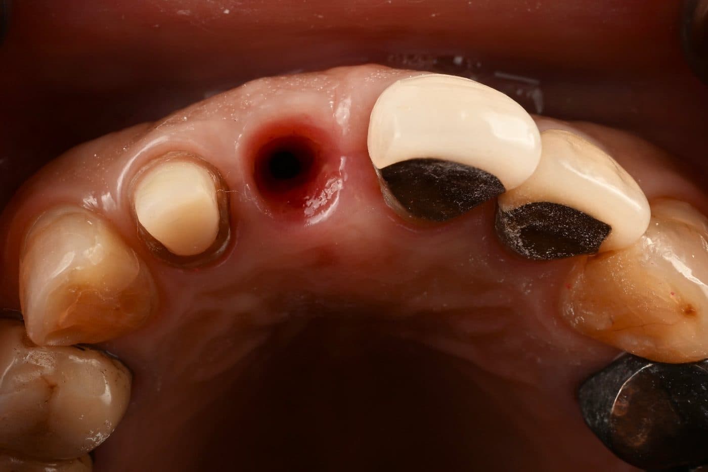
The neighbouring upper lateral incisor (#12) had a crown that no longer fitted well, with decay beneath it. Left untreated, decay under a crown can progress unnoticed, so it was treated at the same time.
After removing the old crown and the decay, the tooth was rebuilt with a fiber post core. A fiber post was chosen over a metal post with the aim of reducing stress concentration on a root that has already had root canal treatment. The tooth was then prepared and restored with a zirconia crown, matched in colour and shape to the new implant crown.
The implant used: Hiossen (Osstem group)
This case used a Hiossen implant, made by the U.S. subsidiary of the Osstem group. The Hiossen implant system is FDA 510(k)-cleared.
According to the manufacturer, the tapered body and thread design are intended to achieve initial stability in soft bone. The surface is SA-treated (sandblasted and acid-etched).
Implant healing generally takes about three months. In a randomized clinical trial comparing three implant surfaces including Osstem SA (early loading in sites healed for at least two months after extraction), stability improved in all groups six weeks after placement, when the definitive restorations were delivered, with no significant difference between surfaces (Körmöczi et al., BMC Oral Health, 2021). Where bone conditions are favourable, an earlier move to the final restoration may therefore be considered for patients who want to shorten treatment. Healing time depends on the bone and on whether grafting is needed; because this case involved bone grafting, about three months was allowed.
Case summary
- Cause: root fracture under a crown placed more than 30 years ago (labial pocket 7 mm with bleeding on probing, no mobility), with labial bone resorption confirmed on 3D CT
- Choice: the neighbouring teeth were also root-canal treated and at higher fracture risk, so an implant was chosen rather than a bridge
- Surgery: no incision; guided extraction, bone grafting (GBR) and implant placement in one surgery (primary stability 30 Ncm); 6 visits in total
- Healing: custom healing abutment for about 3 months; a temporary tooth bonded to the neighbouring teeth maintained appearance
- Final: provisional restoration to confirm appearance and function, then a screw-retained zirconia crown
- Neighbouring tooth: #12 had an ill-fitting crown with decay; rebuilt with a fiber post core and restored with a zirconia crown
- Outcome in this case: the gum contour was preserved without a gum graft (individual results vary)
References
- 公益財団法人8020推進財団「第2回 永久歯の抜歯原因調査 報告書」2018年11月
- Morton D, Wismeijer D, Chen S, et al. Group 5 ITI Consensus Report: Implant placement and loading protocols. Clin Oral Implants Res. 2023;34(Suppl 26):349–356.
- Chen ST, Hamilton A. Immediate implant placement: indications, surgical and prosthetic procedures and outcomes. Forum Implantologicum (ITI). 2019.
- Ruhstorfer M, Güth JF, Stimmelmayr M, et al. Systematic review of peri-implant conditions and aesthetic outcomes of customized versus conventional healing abutments. Int J Implant Dent. 2024;10:61.
- Körmöczi K, Komlós G, Papócsi P, et al. The early loading of different surface-modified implants: a randomized clinical trial. BMC Oral Health. 2021;21:207.
- Hiossen. ETIII SA — product information (manufacturer).
This describes the course of one patient's treatment, published with written consent. Treatment choices depend on each patient's diagnosis, and results vary between individuals. Literature is cited as general background and does not guarantee any outcome.
Treatment, cost and duration
- Patient profile
- Male, 60s
- Diagnosis
- Root fracture of a previously root-canal-treated upper central incisor (#11), crowned more than 30 years ago, with resorption of the labial (front) bone wall — confirmed on 3D CT. At the first examination: labial probing depth 7 mm with bleeding on probing, no mobility. Non-restorable; extraction and replacement indicated. The neighbouring upper lateral incisor (#12) had an ill-fitting crown with decay beneath it.
- Treatment
- Flapless (no incision) extraction of #11 with guided immediate implant placement (Hiossen, CBCT-planned surgical guide) and simultaneous guided bone regeneration (GBR) for the resorbed labial bone, in a single surgery. Insertion torque 30 Ncm; the implant was not loaded. A custom healing abutment shaped to the original gum contour was left for approximately 3 months, while a temporary tooth bonded to the neighbouring teeth maintained appearance. An implant-supported provisional restoration was then used to confirm appearance and function before the final screw-retained zirconia crown was placed. No gum graft was required. #12: the old crown was removed and the decay treated; the tooth was rebuilt with a fiber post core, prepared, and restored with a zirconia crown.
- Material
- Hiossen titanium implant (Osstem group; FDA 510(k)-cleared system) and abutment; custom healing abutment; screw-retained single-layered zirconia crown; bone graft material and membrane (GBR). #12: fiber post core and single-layered zirconia crown
- Period
- Approx. 5 months (approx. 3 months of healing, then a provisional phase before the final crown)
- Visits
- 6 visits in total over ~5 months, including 1 surgery (extraction, bone graft and implant placement on the same day)
- Cost
- Self-pay total ¥1,079,300 (incl. tax). Breakdown — #11: extraction ¥99,900 + implant body ¥299,900 + bone grafting (GBR) ¥189,900 + implant abutment ¥99,900 + zirconia crown ¥179,900 (subtotal ¥869,500); #12: fiber post core ¥29,900 + single-layered zirconia crown ¥179,900 (subtotal ¥209,800). CT scanning, examination and diagnosis, local anaesthesia, the surgical guide, the custom healing abutment, both temporary teeth (the bonded temporary and the implant provisional restoration), and the precision impression are included.
Main risks & side effects
- Implant placement is a surgical procedure; post-operative swelling, bruising, and discomfort may occur.
- In rare cases osseointegration (bone-implant fusion) may not proceed as expected, potentially requiring removal of the implant.
- GBR (bone regeneration) uses bone graft material and a membrane; healing times vary between individuals.
- Immediate placement in the front of the mouth carries a risk of gum recession, which can be higher where the labial bone is thin or damaged. Gum shape varies between individuals, and a gum graft may be needed in other cases.
- A temporary tooth bonded to the neighbouring teeth is used during healing; it can come loose and may need re-bonding.
- #12 is a root-canal-treated tooth restored with a post core and crown; root fracture, recurrent decay, or loosening of the crown may occur in the future.
- Smoking and systemic conditions (e.g. diabetes) may affect the outcome.
- The final crown is placed after approximately 3 months of healing and a provisional phase; this case required 6 visits in total.
- Prosthetic components have a finite lifespan and may need maintenance or replacement in the future.
- This is an elective, privately billed (self-pay) treatment, not covered by Japanese national health insurance.
Self-pay (non-insured) treatment. Individual results vary. Published with the patient's written consent.
Computer-guided implant surgery at RODIN
Planned in 3D, before any incision
A dental implant replaces the root of a missing tooth with a titanium post that fuses with the bone, supporting a crown that looks and works much like a natural tooth. The single biggest factor in a smooth, predictable result is planning — knowing exactly where the implant should go before surgery begins.
Every implant at RODIN is planned on a cone-beam CT (CBCT) scan that shows the bone, the nerve canals, and the sinus in three dimensions. We combine that with a digital scan of your teeth and plan the implant position from the final crown backward — so the implant is placed where the tooth needs it, not just where bone happens to be.
A 3D-printed surgical guide
From that plan we design and 3D-print a surgical guide — a custom template that fits over the teeth or gum and directs each implant to the planned position, angle, and depth.
The workflow is: CBCT scan → digital plan → 3D-printed guide → guided placement. Because the position is decided on the computer and then transferred precisely to the mouth, the surgery itself becomes shorter, calmer, and more predictable than free-hand placement.
Why guided surgery
Guided placement helps the implant avoid the nerve and sinus and land exactly where the final crown needs it — supporting predictable, less-invasive surgery planned around your anatomy before any incision is made.
- Helps avoid the nerve canal and sinus
- Places the implant where the crown needs it (restoration-driven)
- Supports shorter, less-invasive, more predictable surgery
- The plan is reviewed before surgery, not improvised during it
Materials & systems
RODIN primarily uses American-made Hiossen implants (FDA 510(k)-cleared), with Straumann and Nobel Biocare available as premium alternatives. The final restoration is typically a screw-retained zirconia crown, which can be retrieved for maintenance if ever needed.
Implant brand matters for long-term parts availability and documented track record: Straumann implants, for example, have documented success rates above 95% at 10 years in peer-reviewed studies. We confirm the system and the full cost in writing before treatment.
Healing & the timeline for visitors
After placement, osseointegration — the fusion of bone to the implant — typically takes about 3 months before the final crown is fitted; a temporary may be worn in the meantime. Where bone is deficient, grafting can be done, which can extend the timeline.
International patients usually plan two trips: one for placement and one for the final crown, with healing in between. We help sequence the visits around your travel.
General, evidence-based information — not a guarantee of outcome. Individual results vary; suitability is assessed individually.
Frequently asked questions
My front crown feels as if it has shifted. Could it be a root fracture?
It is one possibility. Other causes include a loose post or core, deteriorated cement, or gum disease. The cause is confirmed with an examination that includes measuring the gum pockets (a deep pocket in one spot can suggest a fracture) and, where needed, a 3D CT scan. Seeing a dentist early may allow treatment before more bone is lost.
Can a tooth with a fractured root be saved?
It depends on how and where the root has fractured and on the condition of the surrounding bone. An attempt to keep the tooth is sometimes possible, but when the crack is deep and bone has been lost, extraction is usually recommended.
Are root-canal-treated teeth more likely to fracture?
They are generally considered to be at higher risk, because less tooth structure remains and a post is often placed. Japan's 2018 national survey on the causes of tooth extraction (8020 Promotion Foundation) reported a markedly higher proportion of fractures in teeth without a living pulp.
Can an implant be placed on the same day as the extraction?
Yes, when the bone allows (immediate implant placement). In this case, bone grafting was carried out in the same surgery. When conditions are not suitable, the implant is placed after a healing period following extraction. Suitability is assessed on CT.
Will I have a gap at the front while the implant heals?
In this case, a temporary tooth bonded to the neighbouring teeth was worn throughout healing, so there was no visible gap. The type of temporary depends on the case and is planned in advance.
How can gum recession be avoided with a front-tooth implant?
Assessing the bone on CT beforehand, bone grafting where needed, avoiding incisions where possible, and shaping the gum with a custom healing abutment and a provisional restoration all help. Results still vary, and some cases need a gum graft.
With immediate placement and bone grafting, how long until the final tooth?
Implant healing generally takes about three months, and it tends to be longer when bone grafting is involved. This case took about five months in total, including the provisional phase — longer than the general guide shown in the implant FAQ below. The timeline depends on the condition of the bone.
Is implant surgery painful?
Placement is done under local anaesthesia, and IV sedation by a licensed anaesthesiologist is available for anxious patients or longer surgery. Post-operative swelling and discomfort are expected for a few days and managed with standard pain relief.
How long does the whole process take?
Osseointegration typically takes about 3 months before the final crown is placed; a temporary may be worn in the meantime. International patients usually plan two trips.
How is guided surgery different from free-hand?
With guided surgery the position, angle, and depth are decided on the computer from your CBCT and transferred to the mouth via a 3D-printed guide, rather than judged by eye during surgery. It supports more predictable, less-invasive placement.
Am I a candidate for implants?
Most healthy adults with adequate bone are candidates; bone grafting can address some deficits. Smoking and conditions such as uncontrolled diabetes may affect outcomes. Suitability is assessed on CBCT at consultation.
How long do dental implants last?
Implants are designed for the long term; peer-reviewed studies report success rates above 95% at 10 years for established systems such as Straumann. Longevity depends on hygiene, the health of the gums and bone, and not smoking — and the crown on top may need maintenance over time.
Next step
Have a similar concern?
Book a free online consultation and our English-speaking team will outline a treatment plan and a clear written estimate for your case.
Related treatments
Other case studies
Related reading
Article
Implants vs Bridge vs Denture: How to Choose →
Article
Bone Grafting and Sinus Lifts for Dental Implants in Japan: A Guide for International Patients →
Article
Osstem vs Straumann vs Nobel Biocare: Implants Compared →
Article
Dental Implant Timeline in Japan | Step by Step →
Article
Missing Tooth? Replacement Options Compared →